
Presentation
Has presented after accidentally swallowing a false tooth. X - rays required to check the position of the foreign body.
Patient Data
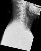
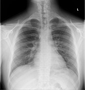

1cm linear foreign body just to the left of the midline at the level of the sternoclavicular joint.
This is lateral to the tracheal air shadow and therefore likely to be in the aerodigestive tract.
Heart size normal. Lungs clear.

Emergency procedure for removal of foreign body. Noted esophageal perforation at 25 cm. Review for biLat pneumothoraces?
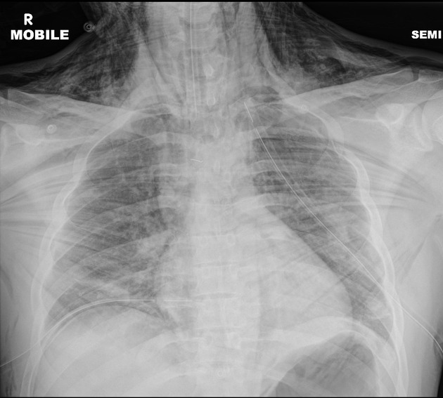
AP
Massive left sided pneumothorax with mediastinal shift.
Moderate right sided pneumothorax.
Pneumomediastinum and pneumoperitoneum.
BL pneumothorax post chest drain insertion
Endotracheal tube. Bilateral pleural drains. Pneumothoraces resolved.
Pneumomediastinum and pneumoperitoneum resolved.
Surgical emphysema with ginkgo leaf sign.
1 cm linear foreign body at T3 level in the midline.
Case Discussion
The first chest radiograph is dramatic with air in the pleural spaces, soft tissues, mediastinum and peritoneal cavity.
The improvement following bilateral pleural drain insertion is equally dramatic having been undertaken less than 1 hour later.


 Unable to process the form. Check for errors and try again.