
Presentation
Headaches and personality change.
Patient Data
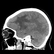

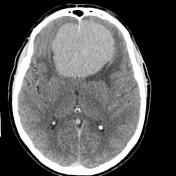

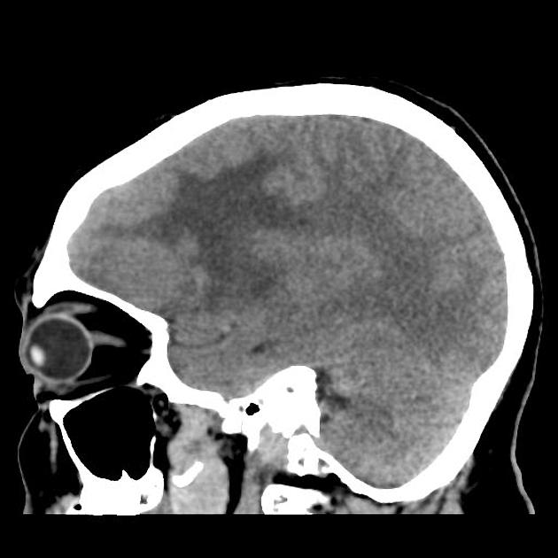
A large hyperdense mass is present in the anterior cranial fossa, appearing to be extra-axial with a broad dural base of contact along the floor of the anterior cranial fossa and the olfactory grooves. Vivid homogeneous contrast enhancement.
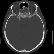
Prominent hyperostosis is present as well as direct extension into the upper parts of the nasal cavity, between the middle ethmoidal air cells.
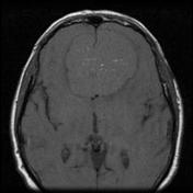
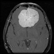
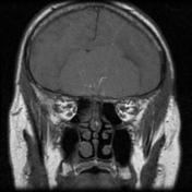
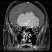
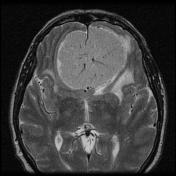
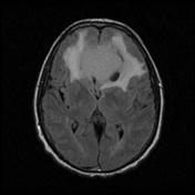
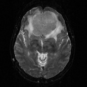
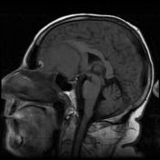

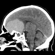
A large lesion, isointense to grey matter on T1 and slightly hyperintense on T2, centered in the anterior cranial fossa. The lesion shows vivid homogeneous enhancement, with a central spoke-wheel of vessels seen arising from the floor of the anterior cranial fossa. CSF cleft with pial vessel noted. The tumor effaces the frontal lobes, frontal horns of the lateral ventricles and the genu of the corpus callosum. There is extensive white matter T2/FLAIR high signal in the frontal lobes, consistent with edema. The lesion wraps around the anterior falx, but is larger on the left than the right.
Enhancement in the roof of the left ethmoid sinuses, with loss of definition of the overlying cortex, is concerning for tumor invasion. The posteroinferior aspect of the tumor extends into the pituitary fossa, separate from the pituitary gland, abutting the medial aspects of both supraclinoid internal carotid arteries. The anterior cerebral arteries extend over the posterosuperior surface of the tumor for much of its extent and are deviated to the right.
A smaller T2 hyperintense lesion at the left posterolateral aspect of the tumor suppresses on FLAIR, consistent with a cyst (such as a peritumoral cyst).
Conclusion
The large frontal meningioma appears to be left olfactory groove in origin, with extension into the pituitary fossa. There is possible invasion into the left ethmoid sinuses, and if clinical relevant fine slice bony CT may be useful to confirm this. There is close approximation of the tumor to both the supraclinoid ICAs and the ACAs.
Case Discussion
The patient went on to have a resection, confirming the diagnosis.
Histology
The sections show a moderately cellular meningioma with attached dura. The tumor forms whorls. No sheeting arrangement is seen. The tumor cells have ovoid nuclei with no nuclear pleomorphism. Mitoses are inconspicuous. There is no necrosis. A small amount of brain parenchyma is seen and there is no evidence of brain invasion. No evidence of atypical or malignant change is identified.
Final diagnosis
Meningioma (WHO Grade I).


 Unable to process the form. Check for errors and try again.