Presentation
Seizure
Patient Data


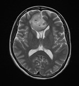
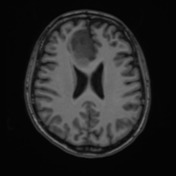
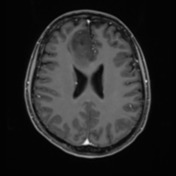
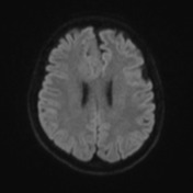
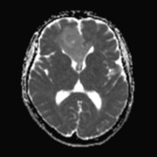
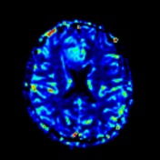
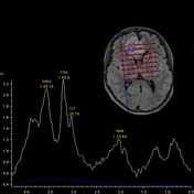
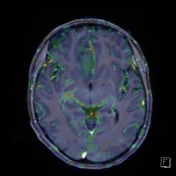

Large expansile FLAIR hyperintense minimally centrally enhancing tumor in the medial aspect of the right frontal lobe centered in the anterior of the cingulate gyrus, involving the genu of the corpus callosum and extending across the midline to involve the medial aspect of the left frontal lobe. The tumor also extends more anteriorly and inferiorly in the frontal lobe to involve the straight gyrus and also inferolaterally along the base of the brain. There are regions of elevated CBV within the tumor. Spectroscopy demonstrates increased choline with reduced NAA but no large lactate peak.
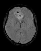
Multiple regions of susceptibility artefact and hypointense on phase imaging within the tumor likely represent calcifications. Midline shift anteriorly measures up to 10 mm displacement of the anterior cerebral arteries to the left
Conclusion:
Large right frontal tumor with subtle central enhancement compatible with infiltrative glioma. Elevated CBV within the tumor may be due to oligodendroglial histology of the tumor rather than indicating a higher grade.
Case Discussion
The patient went on to have a resection.
Histology
Sections show a focally calcified, hypercellular tumor composed of moderately pleomorphic cells containing round to oval, hyperchromatic, vesicular nuclei with conspicuous nucleoli and prominent perinuclear clearing. Tumor cells are arranged in diffuse sheets intersected by thin-walled "chicken-wire" capillaries. Occasional mitotic figures are identified (up to 1/2ohpf). No necrosis or microvascular proliferation is seen.
IMMUNOHISTOCHEMISTRY:
- GFAP: Negative
- NogoA: Positive
- Nesfin: Positive (intermediate)
- IDH-l R132H: Positive (mutated)
- ATRX: Positive (not mutated)
- p53: Positive p16: Positive
- Topoisomerase labeling index: Approximately 5%
Combined 1p/19q co-deletion detected by FISH
FINAL DIAGNOSIS: Oligodendroglioma (WHO Grade II).


 Unable to process the form. Check for errors and try again.