
Presentation
2nd trimester anomaly scan at 21 weeks of gestation.
Patient Data
Gender: Unknown
From the case:
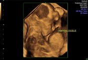
Omphalocele

Download
Info
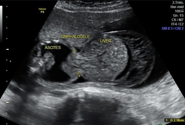
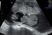
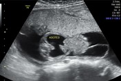
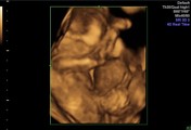
2D/4D ultrasound images of a male fetus (21 weeks gestational age) revealing large mid-line anterior abdominal wall defect with herniating large membrane covered sac containing fluid, multiple echogenic bowel loops as well as the liver & GB. There are associated ascites and marked thickening and edema of the covering membranes.
Findings are highly suggestive of omphalocele.
Case Discussion
The most common differential diagnosis is gastroschisis which is:
- usually smaller para-umbilical defect (usually to the right of the mid-line)
- not covered by membranes
- usually containing only bowel loops


 Unable to process the form. Check for errors and try again.