
Presentation
Painless left side progressive proptosis and conjunctival congestion.
Patient Data


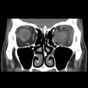
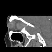
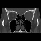
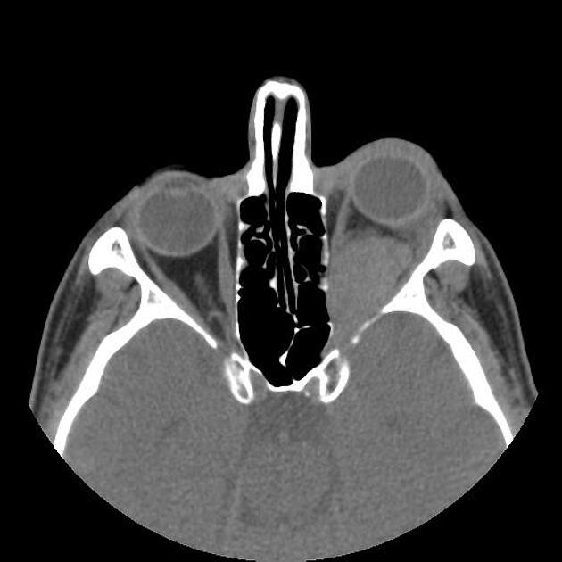
Well-defined lobulated margin solid mass lesion axial width up to 30 x 22 mm and height up to 25 mm in left orbital cavity intraconal middle to posterior part with impression on the adjacent orbital medial wall, related extraocular muscles, the posterior uveo-scleral layer of the eye, and posterior margin of lacrimal gland orbital part are seen. The mass has completely encased the optic nerve sheath complex, leading to left side proptosis, lacrimal gland, proptosis, and lacrimal gland and preseptal soft tissue swelling. Minimal extension of the mass within the optic canal distal segment seems to be also present.
Case Discussion
The case is a pathology-proved optic nerve sheath meningioma with typical features on non-contrast orbital MDCT. The meningioma of the optic nerve sheath is relatively rare and has slow and progressive growth which can lead to optic nerve atrophy, ischemic retina, and blindness. The main treatment of the tumor is surgical excision which because of its proximity to the optic nerve has considerable morbidity. Radiotherapy has increasing role in the management oft he tumor.


 Unable to process the form. Check for errors and try again.