
Presentation
Known prostate malignancy with rapidly rising PSA. CT and bone scan staging prior to chemotherapy.
Patient Data
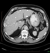


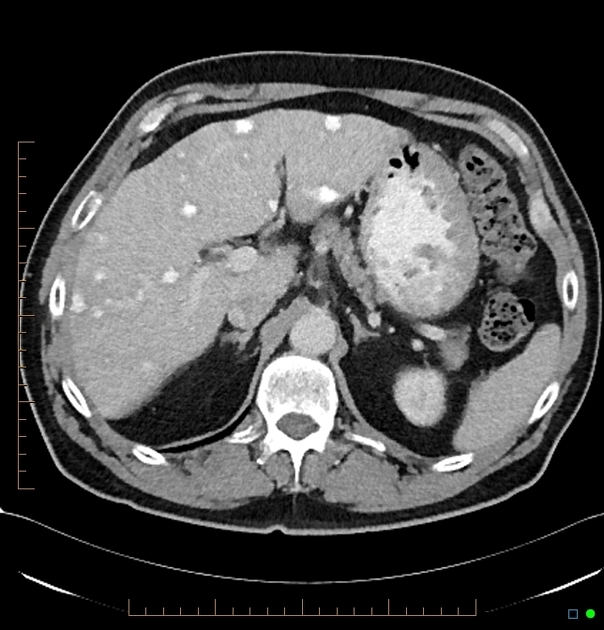
Multiple hyperdense masses throughout the liver with low density rims.
The differential is between calcification and contrast enhancement.

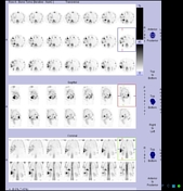
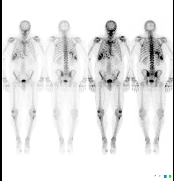
Patchy accumulation of tracer in the liver at the site of hyperdense masses on CT (confirmed on the SPECT imaging) indicating the presence of osteoclasts. Note no abnormal isotope accumulation in the bones.
Case Discussion
Calcified liver metastases are reasonably uncommon and usually associated with metastatic mucinous adenocarcinoma (gastric or colonic in origin). Prostatic metastases are typically sclerotic in bone, but very rarely calcified in soft tissues although it has been reported (see below). Given the positivity on nuclear bone scan, these lesions are not just calcified but actively ossifying. The effect is a primary phenomenon and not post-chemotherapy as the patient has not yet received any such treatment.


 Unable to process the form. Check for errors and try again.