
Presentation
14 year old with knee pain following an injury while playing football.
Patient Data

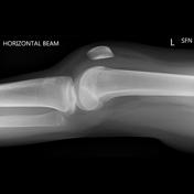
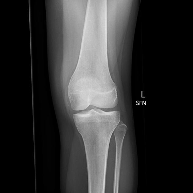
There is a large knee effusion and a bone fragment in the joint. No fracture demonstrated. The effusion and bone fragment are highly suggesting of an ACL injury.
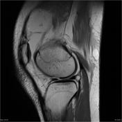

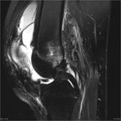
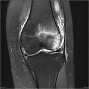
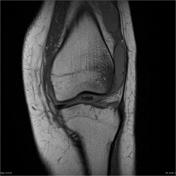
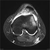

Large osteochondral defect of the femoral condyle and medial patella with loose bodies in the joint. Kissing-contusions secondary to patella dislocation and a shallow trochlear groove. Large joint effusion.
Case Discussion
A serious knee injury with large osteochondral defect of the femoral condyle and medial patella with loose bodies in the joint. There are kissing-contusions secondary to patella dislocation and a shallow trochlear groove, which has predisposed to patella dislocation during the trauma.
On the initial plain film, there is a fragment of bone adjacent to the tibial spine and a large knee effusion. It would be reasonable to presume that this was a result of an ACL injury. But, it was a fragment of bone from the osteochondral defect and there is no ACL injury.
Its also worth noting that a large effusion juxtaposed to the pad underneath the quadriceps tendon can look like a fat-fluid level. In this case, you would be forgiven for describing a lipohemarthrosis on the lateral radiograph, but there is no fat demonstrated at MRI.


 Unable to process the form. Check for errors and try again.