
Note: This case has been tagged as "legacy" as it no longer meets image preparation and/or other case publication guidelines.
From the case:
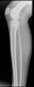
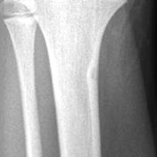
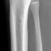
Osteoid osteoma

Download
Info
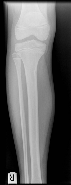
Subtle cortical thickening with a small well defined lucent nidus.
Case Discussion
Incidental osteoid osteoma.


 Unable to process the form. Check for errors and try again.