
Presentation
Reluctance to weight bear on the left leg. Generally unwell with low grade fever.
Patient Data

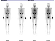
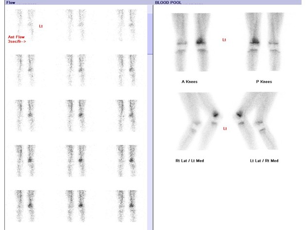
On early phase imaging, there is increased blood flow to the left leg, with particular hyperemia in the region of the distal femoral metaphysis.
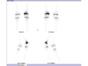
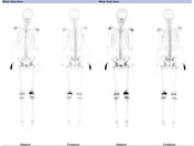
On delayed phase imaging, there is intense tracer accumulation in the corresponding left distal femoral metaphysis, most marked anteromedially. The left distal femoral physis also demonstrates more intense tracer accumulation compared to the left.
Tracer distribution elsewhere is physiological only.
Case Discussion
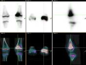
In pediatric patients, the metaphyses of long bones are a common site for osteomyelitis secondary to the increased vascularity to this region in developing bone. On bone scan, osteomyelitis is typically 'three phase positive' with increased blood flow and hyperemia on early phase imaging, and intense increased tracer accumulation (osteoblastic activity) on delayed phase imaging. SPECT/CT is useful for anatomical localization, improving the specificity of nuclear medicine bone scan.
Physes will demonstrate linear increased tracer accumulation, which is physiological. Assessing for assymetry between sides is useful when localizing pathology. A traumatised or infected growth plate will usually demonstrate increased osteoblastic activity compared to the normal contralateral side.


 Unable to process the form. Check for errors and try again.