
Note: This case has been tagged as "legacy" as it no longer meets image preparation and/or other case publication guidelines.
From the case:
Osteomyelitis - proximal tibia

Download
Info
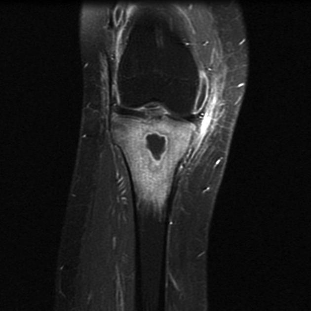
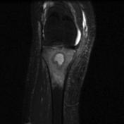
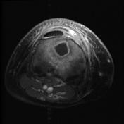
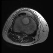
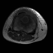
A rounded intramedullary lesion is noted at the proximal metaphysis of the tibia with geographic margins, liquified core, and surrounded by moderate perifocal edema. Lesion margins show post-contrast enhancement. Moderate subcutaneous edema at the anterior aspect of the tibia with underlying cortical interruptions.
From the case:
Osteomyelitis - proximal tibia

Download
Info
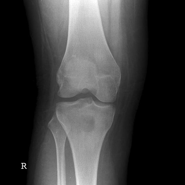
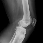
Frontal and lateral views show a metaphyseal rounded lucency at the proximal tibia.
Download
Info
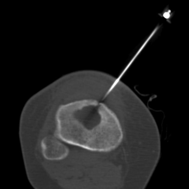
CT-guided biopsy of the lesion.
Case Discussion
Osteomyelitis of the proximal tibia.


 Unable to process the form. Check for errors and try again.