
Presentation
Assesment following road traffic accident.
Patient Data

An expiratory film with magnified heart size.
Clear both lung fields and costophrenic angles.
Linear fracture is seen in the posterior aspect of the left first rib.
Multiple bones islands are seen in both proximal humerus and glenoids suggesting osteopoikilosis.
No evidence of bony injury.
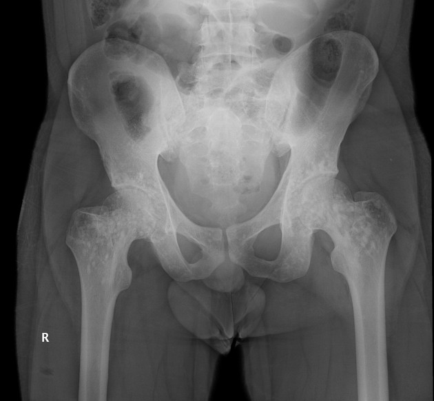
Multiple bone islands are seen in diffuse symmetrical pattern concentrated around the acetabulum, in the pelvic bones and proximal part of both femurs suggesting osteopoikilosis.


Fractures of the posterior portions of the left ninth and tenth ribs are seen.
Fracture of the posterior aspect of the right tenth rib is also seen.
Multiple rounded densities similar to cortical bone are seen throughout the cancellous bone due to osteopoikilosis.
Mild bilateral pleural thickening is seen.
A transitional lumbosacral vertebra is seen with pseudoarthrosis on the left side.
Case Discussion
This is a 25-year-old male presenting through the A&E department after a road traffic accident, urgent ultrasound was requested did not demonstrate suspicious findings of intra-abdominal or pelvic injury. Followed by x-rays and CT scan as requested by the emergency room physician, demonstrated rib fractures, and incidental dense bone within the medullary space with spiculated margins blending within the surrounding trabeculae paralleling them. They appear linear and spherical in shape involving the appendicular skeleton, pelvis and clustered around joints not exceeding 1 cm in size. The patient denied any other symptoms or history of primary tumor. These sclerotic lesions present multiple bone islands (enostoses), finding are those of osteopoikilosis.


 Unable to process the form. Check for errors and try again.