
Presentation
Incidental finding on routine ultrasound exam.
Patient Data
Age: 35 years
Gender: Female
From the case:
Ovarian fibroma

Download
Info

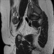
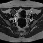
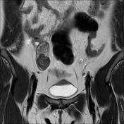
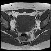
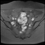
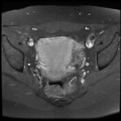
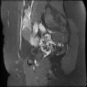
Well-defined right ovarian ovoid mass of isosignal to the myometrium on T1, low signal on T2 with mild enhancement on post-contrast sequences.
Normal appearance of the left ovary containing small follicles.
Mild peritoneal effusion.
Case Discussion
MRI features most consistent with an ovarian fibroma.
On imaging, the main differential diagnosis is thecoma and fibrothecoma which:
most occur in adult women, with ~66% in postmenopausal women
tend to have a brighter signal on T2 given edema and cystic degeneration
contrast-enhancement may be observed given the vascularization of the theca cells


 Unable to process the form. Check for errors and try again.