
Presentation
Pelvic pain
Patient Data
Age: 30 years
Gender: Female
From the case:
Ovarian fibrothecoma

Download
Info

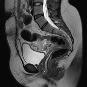
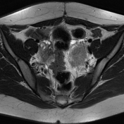
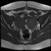
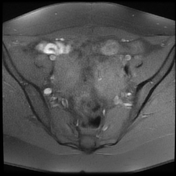
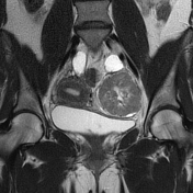
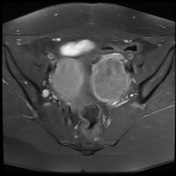
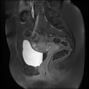
There is a well-circumscribed left centro-ovarian ovoid soft tissue mass of intermediate signal intensity on T1WI, low signal intensity on T2WI (fibrous component) with central high signal areas representing most likely areas of cystic degeneration +/- edema. Mild heterogeneous enhancement is noted on postcontrast sequences. Bilateral hydrosalpinx is noted.
Normal appearance of the uterus.
The right ovary shows normal appearance with small follicles.
Case Discussion
The MRI features are most consistent with an ovarian fibrothecoma
The differential diagnosis should include 1:
- pedunculated leiomyomas
- other solid ovarian masses:
- Brenner tumors
- granulosa cell tumors
- dysgerminomas
- malignant ovarian tumors in the presence of extensive cystic degeneration.p
Additional contributor: A, Ramdani, MD


 Unable to process the form. Check for errors and try again.