Presentation
32/40 pregnant. Right lower quadrant pain.
Patient Data


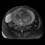

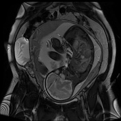


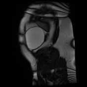

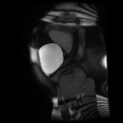

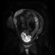

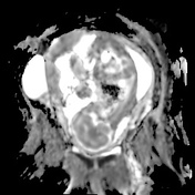

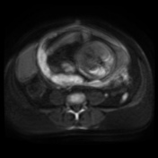

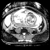

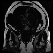

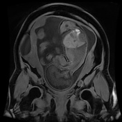

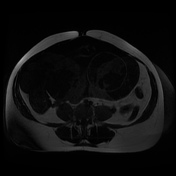


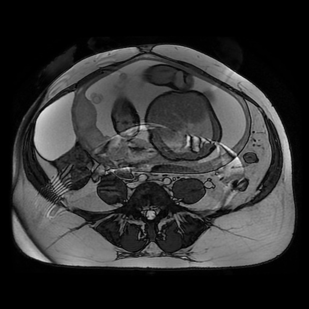
The appendix is not clearly identified, but there is no fat inflammation or restricted diffusion in the region of the cecal pole or elsewhere in the abdomen or pelvis. A unilocular cyst measuring 10 cm in diameter arises from the right ovary. There is no mural irregularity or internal septation, and no intra-lesional hemorrhage. The ovarian vessels are noted to be distended.
Due to its size, the cyst was removed at C-section at term.
Histopathology
Specimen: Right ovarian cyst wall.
Clinical history: C-section + Rt ovarian cystectomy. Large ovarian cyst ~ 10-12 cm. Simple -clear fluid.
Macroscopic: Collapsed cyst 80 x 25 x up to 15 mm. No solid areas.
Microscopic: This specimen consists of a cyst wall lined by benign mucinous epithelium. There is some architectural complexity but there is no cytological atypia or conspicuous mitotic activity in these areas. There is no borderline change or malignancy.
Conclusion: Right ovarian cyst wall - benign mucinous cystadenoma.
Case Discussion
The only abnormality identified was the right ovarian cystic lesion, and the patient progressed to term and the cyst was removed at C-section, with pathology analysis revealing a mucinous cystadenoma.
This study also demonstrates a common finding in pregnancy; the ovarian veins are distended and can be tracked in the retroperitoneum up to the terminations in the IVC on the right and the left renal vein on the left side.


 Unable to process the form. Check for errors and try again.