
Presentation
Chronic abdominal pain.
Patient Data


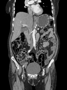
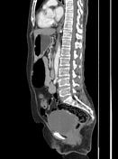
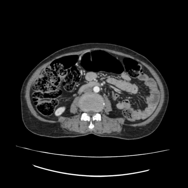
Uncinate process and part of the head of the pancreas are visible. The dorsal portion of the pancreas appears absent. Instead, the retroperitoneal fat replaces the portion of the pancreas lacking. The ducts of Santorini and Wirsung are visible, communicating with minor and major duodenal papillae, respectively. There are multifocal dense calcifications with an irregular cavity in the ventral pancreas, marking the features of chronic pancreatitis.
Case Discussion
No associated abnormal findings in the abdomen were found such as heterotaxi, polysplenia, and renal cysts. There was no mass in the pancreas or features of pancreatic lipomatosis, as the ducts could not be visualized in the agenesis of the dorsal pancreas. The imaging features are characteristic of partial agenesis of the dorsal pancreas. Multifocal calcifications with an irregular small cavity could represent chronic pancreatitis. The pancreatic enzymes were in the normal range. The patient didn’t have any signs or symptoms of diabetes. The HbA1c and random blood glucose levels were within normal range.
Case co-authors:
Dr Farhad Farzam, Kabul University of Medical Sciences
Dr Milad Fikat, Maiwand Teaching Hospital
Dr Javid Karimy, Wyagal Radiology Center
Dr Najibullah Rahil, Kabul University of Medical Sciences


 Unable to process the form. Check for errors and try again.