
Presentation
6 years history of infertility with severe RLQ pain, and tenderness.
Patient Data
Age: 30 years
Gender: Female
Download
Info
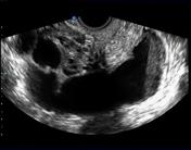
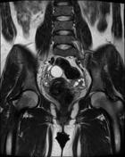
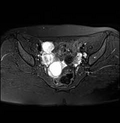
A right adnexal dead end, tubular, fluid filled structure, with typical mucosal infoldings toward the lumen, thick wall and a debris-fluid level (ultrasound image), characteristic of a dilated fallopian tube (in this case: pyosalpinx).
Right ovary is noted medial to the dilated fallopian tube and has normal appearance. Mild free fluid is noted in cul de sac and around the right adnexa.
No accompanying abscess was noted.
From the case:
Pelvic inflammatory disease and pyosalpinx

Download
Info
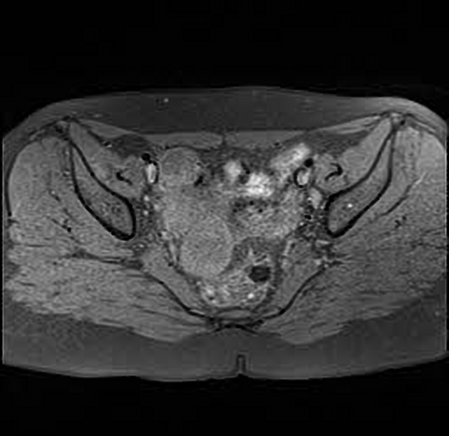
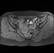
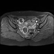
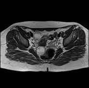
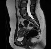
MRI confirmed ultrasound findings. The thick wall of the right fallopian tube is enhanced.
The debris-fluid level is best noted in sagittal T2 images.


 Unable to process the form. Check for errors and try again.