
Presentation
Ventriculoperitoneal shunt inserted after surgery for brain tumor. Routine follow up.
Patient Data


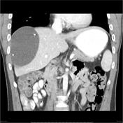
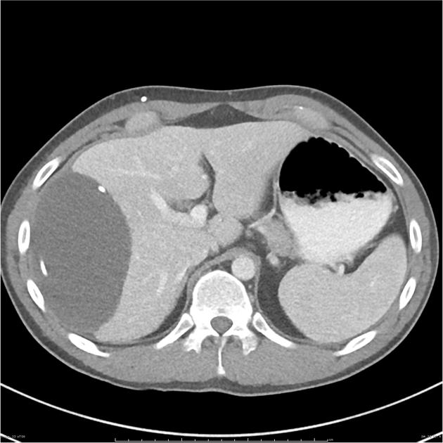
CT of the upper abdomen shows a large water density collection in the right upper quadrant, engulfing the ventriculoperitoneal shunt tubing. The collection exerts mass effect on the adjacent liver. A smaller collection is seen surrounding the shunt in the gallbladder fossa.
The collection had been stable over serial follow-up CT scans spanning 3 years. While biochemical assay of the collection has not been performed at our institution to confirm CSF, the proximity to the shunt makes this most likely.
Case Discussion
This pseudocyst has been described as peritoneal, but it is possible that the VP shunt has penetrated the hepatic capsule, and the large collection may in fact be subcapsular.


 Unable to process the form. Check for errors and try again.