
Presentation
Ataxia, vomiting and papilledema.
Patient Data
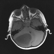

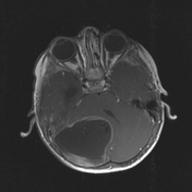
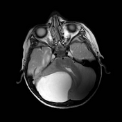
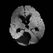
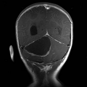
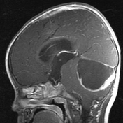

Large cystic mass in the right cerebellar hemisphere with peripheral thick nodular enhancement.
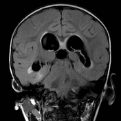
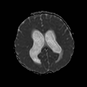
Significant mass effect on the cerebellum and the fourth ventricle, crowded foramen magnum, signs of obstructive hydrocephalus and transependymal edema.
Case Discussion
The patient underwent surgical resection of the tumor.
Histology
Microscopic description: Bi-phasic growth pattern of alternating microcystic and more compact cellular areas. The cells exhibit hair-like processes embedded in a fibrillary background. No evidence of atypia, nuclear pleomorphism, necrosis or increased mitosis is seen. Those glial cells are strongly positive for GFAP immunostatins, Ki-67 proliferative index is estimated at 3%.
Final diagnosis
Pilocytic astrocytoma - WHO grade I.


 Unable to process the form. Check for errors and try again.