Presentation
Two weeks of headaches, nausea and vomiting, drowsiness.
Patient Data
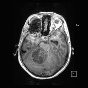

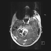


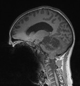

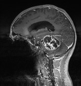

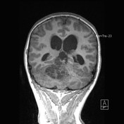

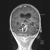

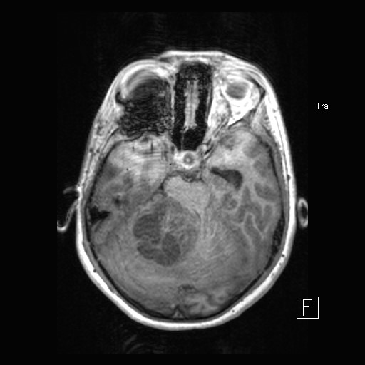
There is a T2 hyperintense, heterogeneously enhancing mass centered on the right cerebellar hemisphere with superior extension into the right ambient cistern measuring 5.0 x 4.7 x. 4.7 cm. There is marked mass effect on the right cerebellum, fourth ventricle, cerebral aqueduct and midbrain/pons, with persistent obstructive hydrocephalus and prominent T2 hyperintensity of the periventricular white matter suggesting transependymal CSF flow. The mass demonstrates multiple foci of susceptibility hypointensity likely representing prominent vasculature. There is moderate surrounding edema involving the right cerebellar hemisphere, and to a lesser degree the right pons, left midbrain, and left cerebellar white matter; mild edema extends into the medulla.
Pathology Report:
In concert with the piloid morphology and clinical/radiological characteristics, the findings are most consistent with a pilocytic astrocytoma.
Case Discussion
The classic appearance of a pilocytic astrocytoma on MRI is T1 iso to hypointensity with vivid contrast enhancement. On T2, the lesion will appear hyperintense with the cystic component having a high signal.
They are a slow-growing, well-circumscribed tumor with a good prognosis (5 and 10 year survival rate >95%).


 Unable to process the form. Check for errors and try again.