
Presentation
Headache.
Patient Data
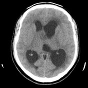

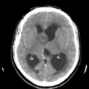

An irregularly enhancing mass centered below the foramina of Munro. No calcification.

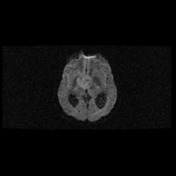
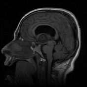
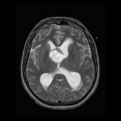
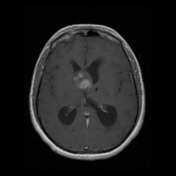
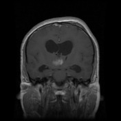
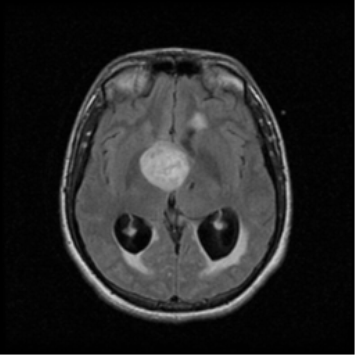
Large intraaxial mass lesion, measuring 3.5 x 2.5 x 2.5cm in perpendicular dimensions, it appears to be within the 3rd ventricle, obstructing the monro foramina bilaterally, causing severe bilateral obstructive hydrocephalus with transependymal edema evident. The mass inferiorly displaces the floor of the third ventricle floor, hypothalamus and mammillary bodies and optic chiasm.
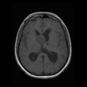
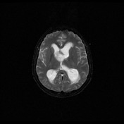
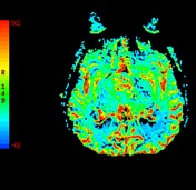
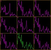
Irregular enhancement. There is no significant surrounding edema, with no significant diffusion restriction (>1200 x 10-6 mm2/s). Decreased NAA, increased Choline and CBV noted within the mass.
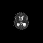
Right frontal ventricular drain in situ, traversing the left lateral ventricle with the tip appears within the left thalamus.
The patient went on to have an endoscopic biopsy.
Histology
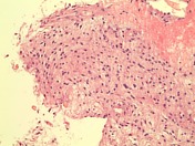
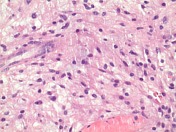
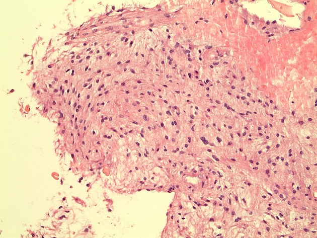
MICROSCOPIC DESCRIPTION:
The sections show features of a moderately cellular glial tumor. Scattered bipolar astrocytes are seen, which show focal somewhat fascicular arrangement. The tumor cells have elongated and hyperchromatic nuclei. No Rosenthal fibers or eosinophilic granular bodies are seen in the background. No mitoses are present. No microvascular proliferation or necrosis is identified. The features are those of pilocytic astrocytoma. The topoisomerase index is less than 1%. IDH-1 immunostain is negative.
DIAGNOSIS: Hypothalamic tumor: Pilocytic astrocytoma (WHO Grade I).
Histology courtesy of Dr Alpha Tsui, Royal Melbourne Hospital.
Case Discussion
Appreciating that this mass is essentially within the third ventricle without surrounding edema or restricted diffusion helps point you towards a low grade tumor. In this age group a pilocytic astrocytoma should be favored.


 Unable to process the form. Check for errors and try again.