
Presentation
Headaches.
Patient Data
Age: Child
Download
Info
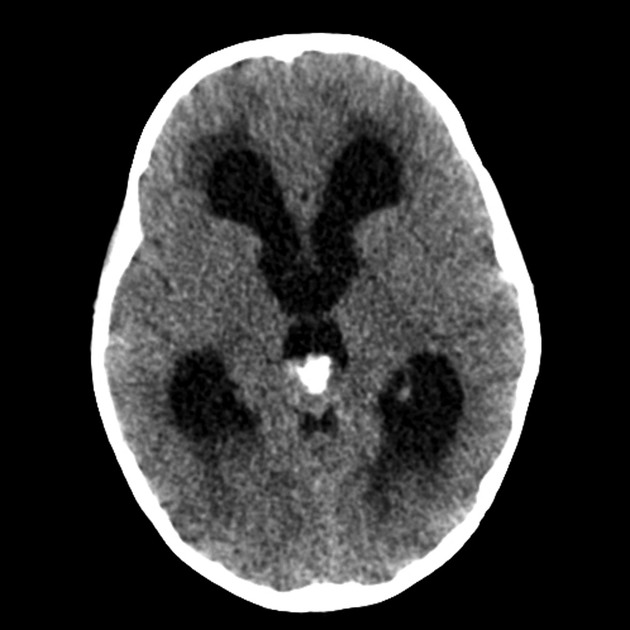
Single image through the brain demonstrates a soft tissue mass in the region of the pineal gland with eccentric calcification (anterior) and evidence of hydrocephalus.


Download
Info
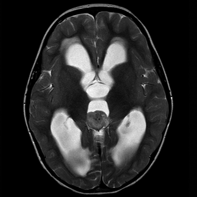
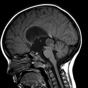
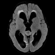
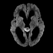
An enhancing mass in the region of the pineal gland is present which demonstrates restricted diffusion on DWI. A tongue of tissue is seen extending inferiorly through the aqueduct, obstructing it and resulting in hydrocephalus with transependymal edema.
Case Discussion
The patient went on to have surgery which confirmed the diagnosis of a pineoblastoma, as wells as confirming extension of the mass down aqueduct.


 Unable to process the form. Check for errors and try again.