
Patient Data
Age: Elderly
Gender: Male
Note: This case has been tagged as "legacy" as it no longer meets image preparation and/or other case publication guidelines.


Download
Info
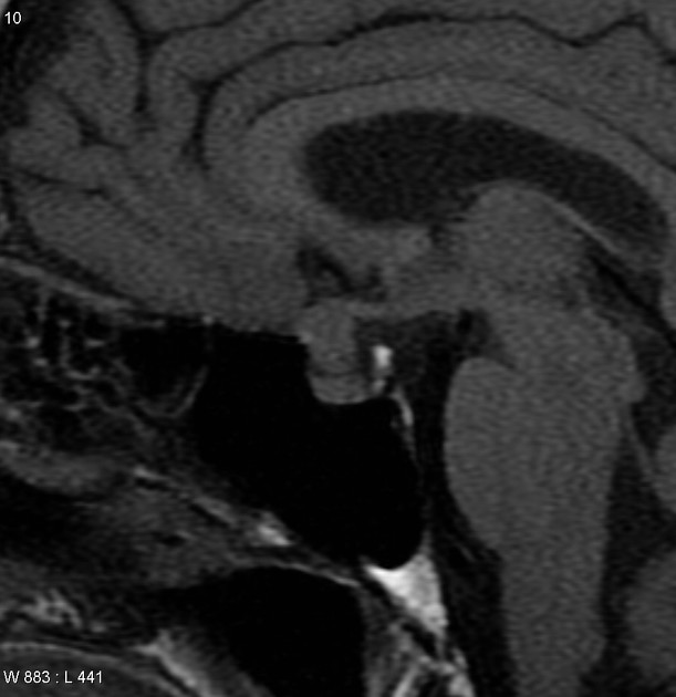
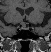
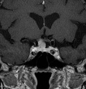
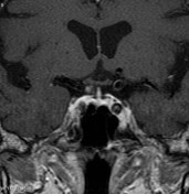
Small right sided enhancing mass with a small dural tail separate from the pituitary gland is most consistent with a meningioma of the anterior wall of the pituitary fossa and planum sphenoidale, compressing and displacing the right optic nerve.


 Unable to process the form. Check for errors and try again.