
Note: This case has been tagged as "legacy" as it no longer meets image preparation and/or other case publication guidelines
From the case:
Pleomorphic adenoma


Download
Info
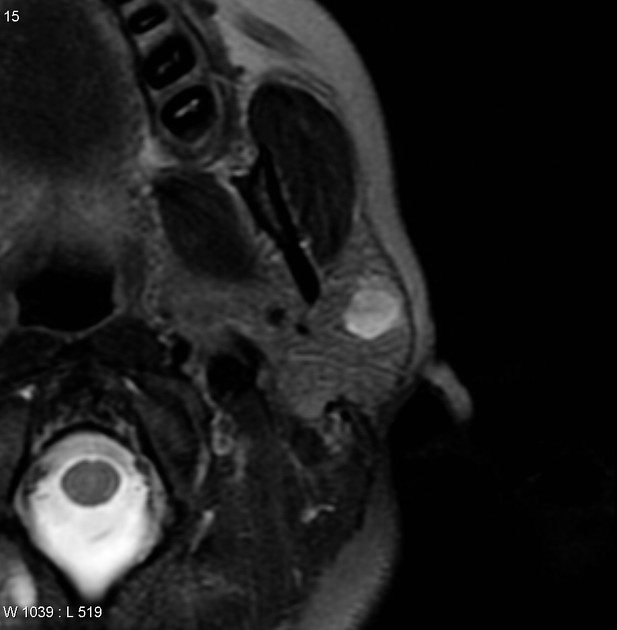
MRI through the left parotid gland demonstrates a sharply circumscribed solid mass in the superficial aspect of the gland. It is isointense to muscle on T1 and strikingly hyperintense on T2. Following contrast administration, it enhances uniformly (note the chemical shift artifact at the margins of the lesion due to the interface between the mass and the intra-parotid fat).
Case Discussion
Features are characteristic of a pleomorphic adenoma which was subsequently excised and confirmed histologically.


 Unable to process the form. Check for errors and try again.