
Presentation
Past medical history of marijuana use and cyclic vomiting syndrome, now presenting with several days of emesis.
Patient Data
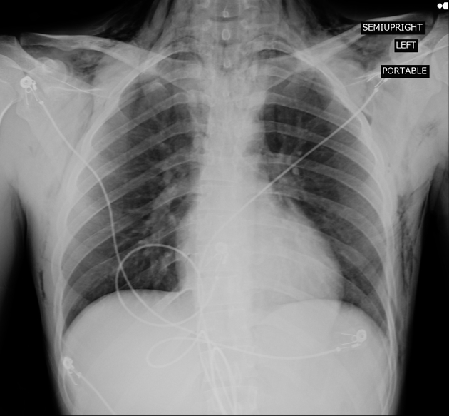
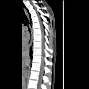
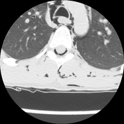
Extensive subcutaneous emphysema in the chest and neck.
No large pneumothorax.
Overlying telemetry leads.
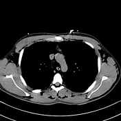

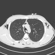
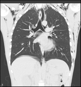
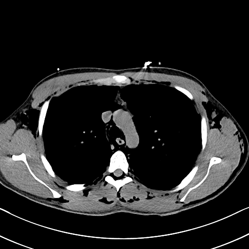
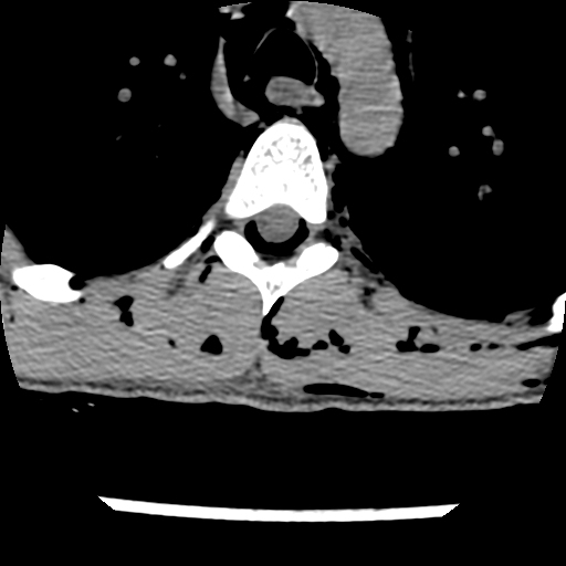
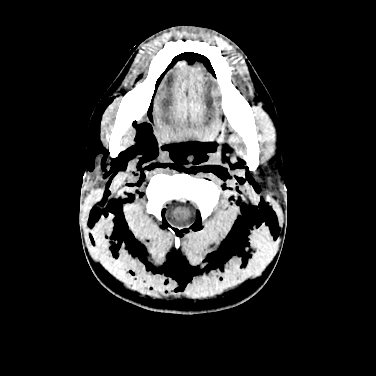
Extensive soft tissue gas in the chest and neck.
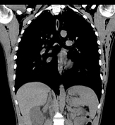
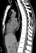
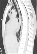
Pneumomediastinum and gas in the extradural space.
No rib fracture.

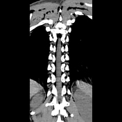
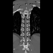
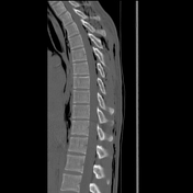
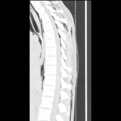
Extensive soft tissue gas in the back.
Partially imaged pneumomediastinum. Gas in the extradural space around the spinal cord, likely extending from the mediastinum via the neural foramina. The gas collects posteriorly rather than anteriorly in the spinal canal.
Tiny bilateral pneumothoraces at the lung apices.
No thoracic spine fracture.
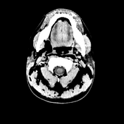
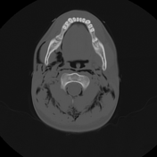
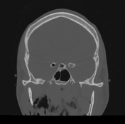
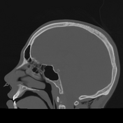
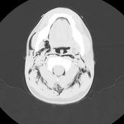
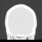
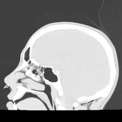
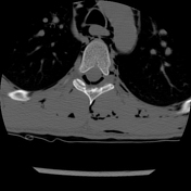
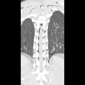
Extensive soft tissue gas in the head and neck. Gas in the extradural spinal canal extending up to the foramen magnum.
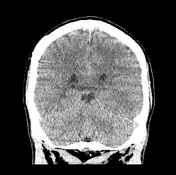
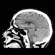
No pneumocephalus.
Involuntary patient motion due to hiccups.
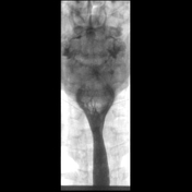
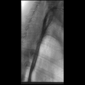
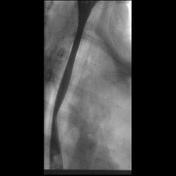

Single contrast oesophagram (cine series) with Gastrografin and subsequently with thin barium demonstrates no extraluminal contrast to suggest esophageal perforation.
Case Discussion
On imaging, this patient was found to have gas collecting in the spinal canal, a condition known as pneumorrhachis. This rare phenomenon typically arises secondary to trauma, surgical procedures, pneumothorax, or pneumomediastinum, and rarely presents spontaneously 1. Notably, as demonstrated in this case, it is crucial to evaluate for pneumorrhachis in marijuana users, given the rising incidence of cannabinoid hyperemesis syndrome 2.
While most cases are asymptomatic, some patients may have neurological symptoms or lumbar and radicular pain 1,3. Differentiating between extradural and intradural pneumorrhachis is important as the clinical implications differ; extradural pneumorrhachis is generally self-limiting, whereas intradural pneumorrhachis can lead to more serious complications such as meningitis and/or pneumocephalus 4.
Treatment is usually conservative, with the gas resorbing spontaneously 4. However, in cases of traumatic pneumorrhachis careful monitoring is necessary as the presence of air suggests severe trauma 4.
Case co-author: Samantha Diulus, MD (Loyola University Medical Center)


 Unable to process the form. Check for errors and try again.