
Presentation
Desaturation and SOB. Background of COPD, glottic SCC and tracheostomy.
Patient Data
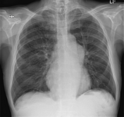
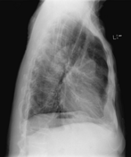

The tracheostomy tube and the left PICC are appropriately positioned. No evidence of consolidation or a pleural effusion.
Enlarged pulmonary trunk. No cardiomegaly.
Left-sided PICC in situ, tip at the RA/SVC junction. Evidence of previous laryngectomy with tracheostomy and TO puncture tube in situ.
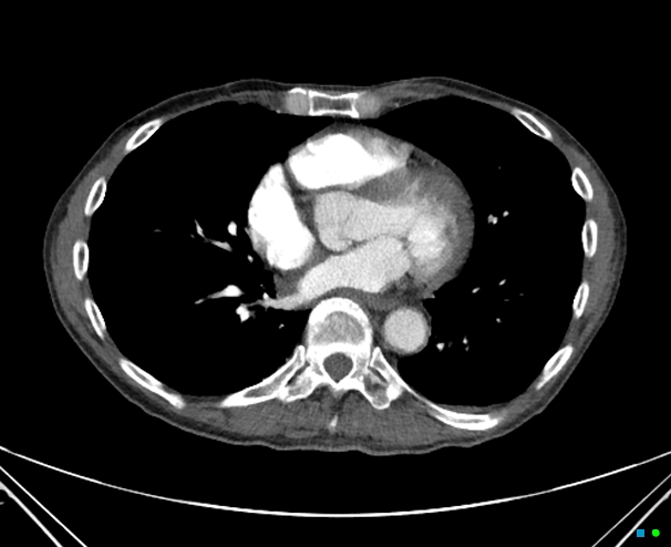
Diagnostic quality CTPA. No filling defect to the subsegmental level. The pulmonary trunk is aneurysmal as are the right and left pulmonary arteries (left > right) which is unchanged from prior imaging. The pulmonary valve has 4 cusps. Small pericardial effusion.
Enlarged mediastinal or hilar lymphadenopathy by CT size criteria.
IMPRESSION
Negative study for pulmonary embolism. Stable pulmonary artery aneurysm.
Case Discussion
The patient had a long history of pulmonary valvular stenosis.


 Unable to process the form. Check for errors and try again.