
Presentation
Admitted to hospital febrile with rigors and found to be in septic shock 4 weeks after an uneventful planned Cesarean section.
Patient Data
On examination there was no apparent source of infection. The surgical wound was clean. No signs of peritonitis but some vague left upper quadrant tenderness. An ultrasound was acquired.
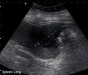
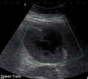

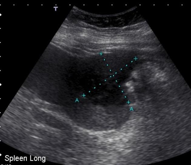
The spleen measures 13.8 cm and demonstrates an irregular hypoechoic lesion in the upper pole measuring 4.6 cm x 4 cm x 4.2 cm. No internal vascularity is seen in this region. The remainder of the scan was normal.


There is a complex cystic lesion in the inferolateral aspect of the spleen with surrounding inflammatory change. Appearances are compatible with an abscess. The adjacent splenic flexure demonstrates bowel wall thickening. The bowel changes may either be primary or secondary to the splenic abscess.
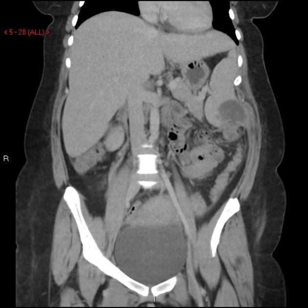
It was decided that due to the location of the cyst and the unilocular nature CT guided drainage was an appropriate treatment choice.
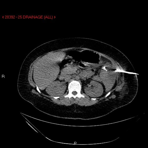
Using an aseptic technique and CT guidance a 15-cm x 18-gauge needle was inserted into the splenic abscess and approximately 10 mLs of a pus filled collection withdrawn. A sample was sent to pathology. The tract was then dilated and an 8.5 French pigtail catheter inserted without complication.
Case Discussion
The specimen grew:
gram negative rods, gram positive cocci
Enterococcus faecalis, Streptococcus milleri and mixed anaerobes
A colonoscopy was also performed which was normal.
A transthoracic echocardiogram was also obtained due to the link between splenic abscess and infective endocarditis, reported as normal.
The patient was treated with IV antibiotics and had the drain removed three days later when draining had stopped. The patient was discharged with no further complications.
No cause for the abscess was found. There may have been some trauma to the spleen during delivery, which was then colonized by bacteria, but this cannot be proven.


 Unable to process the form. Check for errors and try again.