
Presentation
Headache and vomiting
Patient Data


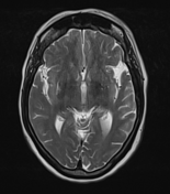
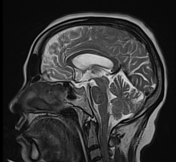
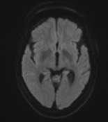
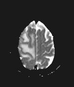
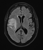
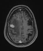
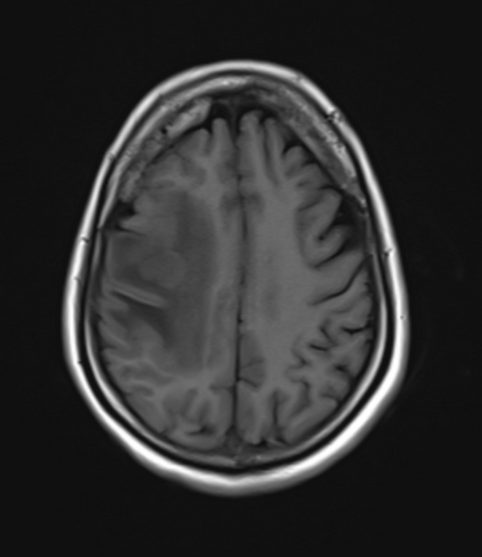
There is a small, well-defined lobulated mass lesion in the right superior frontal lobe in the subcortical region with marked surrounding vasogenic edema. The lesion appears isointense to grey matter on T1W / T2W/FLAIR images and shows diffusion restriction on DWI. Vivid homogenous enhancement is seen on post-contrast images. Another tiny nodular enhancing lesion is seen just posterosuperior to the above lesion.

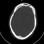
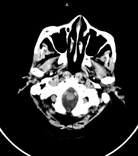
Post-biopsy CT brain shows the lesion is slightly hyperdense with moderate surrounding edema.
Craniotomy defect in the right frontal bone.
Hyperostosis frontalis interna seen ( Insignificant finding).
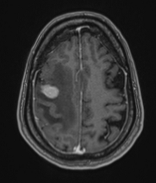
Notch sign refers to irregular growth pattern as well as pliable and infiltrative properties of the tumor.
Case Discussion
The immunohistopathological report of the lesion shows diffuse large B-cell lymphoma (DLBCL). There is no co-existing systemic disease and the patient was immunocompetent.
Co-author: Dr. Deepa N A. DM (Neuro medicine).


 Unable to process the form. Check for errors and try again.