
Presentation
Deterioration in general state, fever, anemia, rise in inflammatory markers and hepatic function tests.
Patient Data


In comparison to previous CT abdomen done almost 2 years previously (see study below):
Small left pleural effusion.
Status post cholecystectomy and hysterectomy.
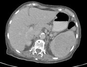
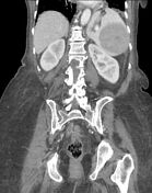
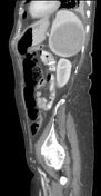
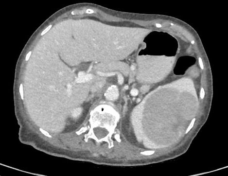
Large hypodense hypoenhancing splenic mass measuring 9.0 x 7.4 x 7.7 cm - has grown considerably.
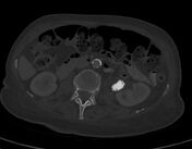
Cystic lesion in body of pancreas measuring 11 x 9 mm - not visible on previous study.
Large jackstone calculus in left extrarenal pelvis - unchanged.
Parapelvic cyst in left kidney.
Tiny amount of free intraperitoneal fluid in pelvis.
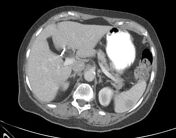
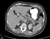

CT abdomen from almost 2 years prior:
The splenic mass was significantly smaller.
The calculus in the left proximal ureter was the same size.

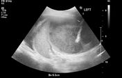
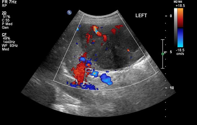
Ultrasound-guided core biopsy obtained from the splenic mass.
Case Discussion
An elderly woman was admitted to the hospital with a febrile illness. CT abdomen showed a large hypodense splenic mass. A previous study from almost 2 years earlier had shown a small splenic mass that had not been investigated further. On the current occasion, a core biopsy was promptly obtained from the mass.
Spleen (core biopsy): diffuse large B-cell lymphoma. Fibrotic core infiltrated by a population of large atypical cells which are positive for CD19, CD20, CD45 (LCA), CD79a, and Bcl6, and negative for CD3, CD5, CD10, CD23, cyclin-D1, and pankeratin. Ki67 index: 70-80%.


 Unable to process the form. Check for errors and try again.