
Presentation
Lower back pain. History of prostate cancer.
Patient Data
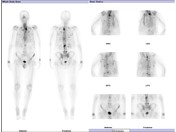
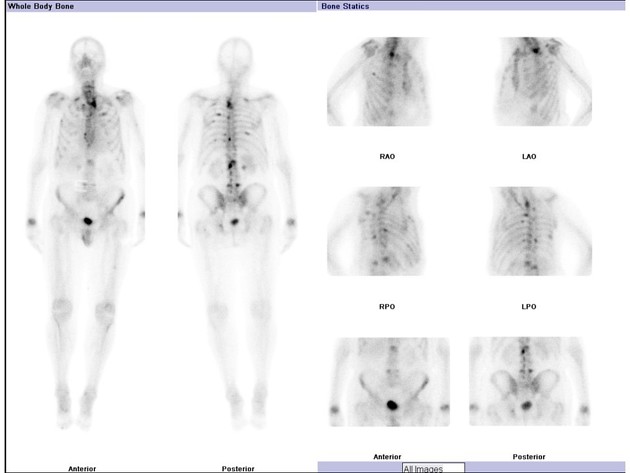
There are multiple foci of increased tracer accumulation throughout the axial and proximal appendicular skeleton. Lesions are most numerous in the thoracolumbar spine and bilateral ribs.
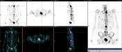
SPECT/CT highlights a large lesion in the upper thoracic spine, where intense increased osteoblastic activity extends into the lateral elements. Multiple additional osteoblastically active lesions are seen on the MIP projection.
Case Discussion
Prostate cancer metastases typically demonstrate increased osteoblastic activity on bone scan. In patients with widespread metastases, such as in this case, lesions are more numerous in the axial and proximal appendicular skeleton (particularly the pelvis). Sclerotic lesions often accompany the osteoblastic lesions on low dose CT.


 Unable to process the form. Check for errors and try again.