Presentation
Mixed stress and urge urinary incontinence. Vaginal discomfort. Difficult defecation.
Patient Data


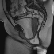
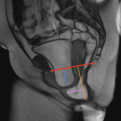
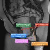
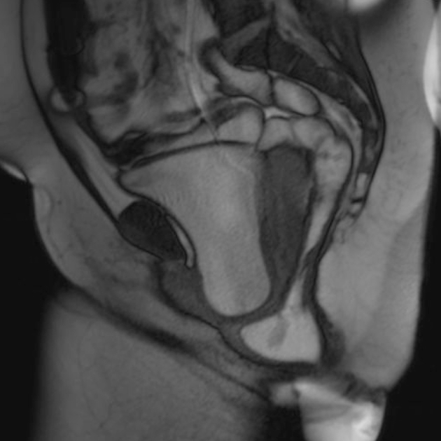
These images show the measurements taken on an MRI proctogram study, and are based on this case.
The measurements are taken at maximal straining and are as follows:
Bladder neck: 37 mm below line (mild cystocele)
Uterine cervix: 12 mm below line (mild uterine descent)
Anorectal junction: 63 mm below line (moderate anorectal junction descent)
Anterior rectocele: 36 mm in depth (moderate sized anterior rectocele)
Case Discussion
The pubococcygeal line is drawn on a mid sagittal image taken from the evacuatory phase of an MRI proctogram study, from the inferior margin of the pubic symphysis, to the final coccygeal segment, and corresponds anatomically to the plane of the levator muscle. Perpendicular lines are drawn from organ specific reference points in the anterior, middle and posterior compartments, and the measurements converted to grades of organ descent using the rule of 3s:
- 0-3 cm: mild descent
- 3-6 cm: moderate descent
- > 6 cm: severe descent
From the anterior and middle compartments, 1 cm is subtracted from the measured distance between the PCL and the organ-specific reference points, to allow for normal downward movement of the organs during straining. For the posterior compartment, 3 cm is subtracted from the measurement before the conversion to a grade, to allow for the normal lower position of the anorectal junction relative to the levator.
For the grading of anterior rectoceles, the rule of 2s is used:
- 0-2 cm: mild rectocele
- 2-4 cm: moderate rectocele
- > 4 cm: large rectocele


 Unable to process the form. Check for errors and try again.