
Presentation
Incidental finding on CT neck
Patient Data


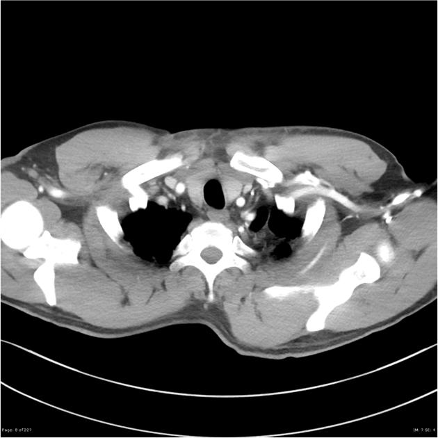
Patient went on to have a formal CT chest for further investigation
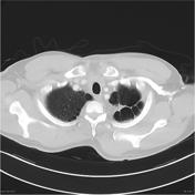
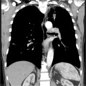
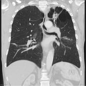
12 x 20 x 36 mm (ML x AP x SI) cavity is again noted within the left upper lobe abutting the oblique fissure and contains a low density nodule with surrounding air. Blebs and bulae are seen throughout the left lung with bullae adjacent to the oblique fissure. Scarring is seen within the left upper lobe and
superior segment of the lower lobe.
Minor pleural thickening associated with scarring in the left costophrenic angle and along the left lateral chest wall. On the right side scarring with associated atelectasis are seen within the posterior subpleural region of the lower lobe and in the lateral superior lower lobe with adjacent ground glass opacity seen associated with this focus of consolidation. Right apical blebs also noted.
Conclusion
Bronchiectasis, scarring and bullae are associated with a left apical cavity containing a soft tissue nodule, is in keeping with a fungal ball (presumably Aspergilloma).
Case Discussion
Typical appearance of a pulmonary aspergilloma.


 Unable to process the form. Check for errors and try again.