
Presentation
Shortness of breath and non-productive cough.
Patient Data


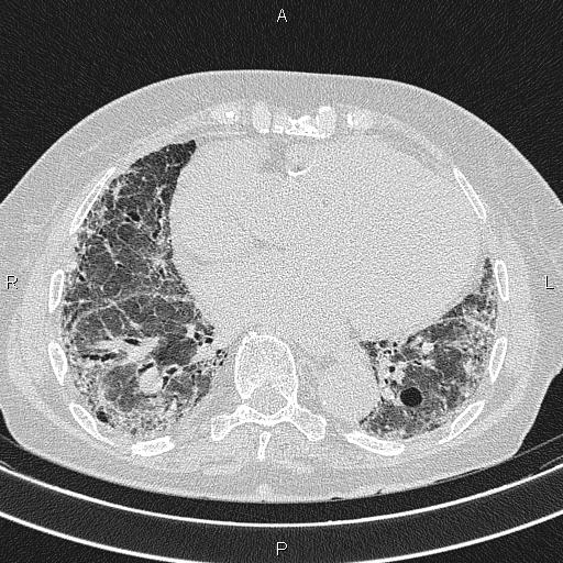
Fibrotic changes are seen in both lungs, including subpleural reticular opacities, honeycombing, tractional bronchiectasis, and parenchymal architectural distortion, which are more prominent in the bases. A few small, thin-walled air cells are also observed bilaterally.
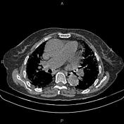
Cardiomegaly is present, and the ascending aorta and the pulmonary artery trunk are dilated. A small volume of pericardial effusion is also seen.
Incidental azygos lobe is evident as an anatomical variant.
Case Discussion
Pulmonary fibrosis can be localized, segmental, or lobar or affect the entire lungs. CT scans can reveal various features that suggest the presence of pulmonary fibrosis, including honeycombing, traction bronchiectasis, lung architectural distortion, reticulation, and interlobular septal thickening.


 Unable to process the form. Check for errors and try again.