
Presentation
Chronic cough.
Patient Data
Age: 25 years
Gender: Female
From the case:
Pulmonary hamartoma


Download
Info
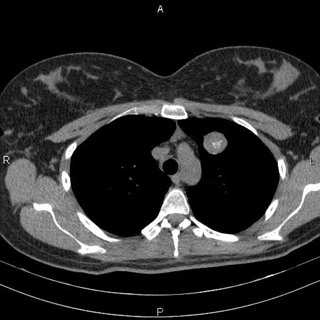
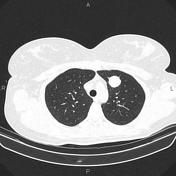
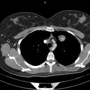
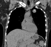
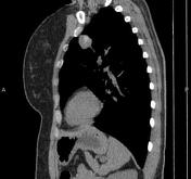
A 27×25mm well defined partially calcified mass is noted at left upper lobe which shows slightly marginal enhancement on post contrast images.
The right subclavian artery is arising from the arch of aorta directly posterior to the esophagus, inferring aberrant right subclavian artery.
A few well defined benign looking masses are seen at both breasts.
Case Discussion
Features are most consistent with pulmonary hamartoma which is one of the most common benign tumors of the lung.


 Unable to process the form. Check for errors and try again.