Presentation
Right upper limb weakness.
Patient Data
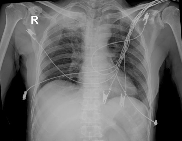
Well-defined round opacity in the right upper zone with an area of relative transradiancy.
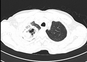

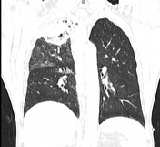

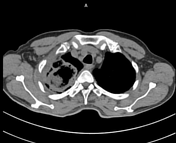

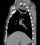

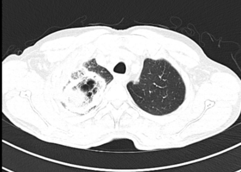
There is a well-defined mass with thick peripheral consolidation (atoll/reversed halo sign) in the apical segment of the right upper lobe with cavitation in the area of relative central transradiancy (bird's nest sign).
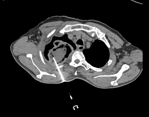
CT guided biopsy from right upper lobe lesion.

Specimen title: CT guided core needle biopsy from right upper lobe lesion.
Gross findings: Received grey-white needle biopsy largest measuring 1 x 0.1 cm.
Microscopic findings: Sections show pulmonary tissue. Alveoli are filled with acute inflammatory cell exudate admixed with fibrin. Also noted are multiple broad hyphae of the mucosa.
Impression: Mucormycosis with acute pneumonitis.
Case Discussion
Initially it was thought that his right upper limb weakness was due to a stroke, however both a CT brain and a subsequent MRI brain were normal. The routine CXR revealed an unexpected right apical mass and CT demonstrated the typical bird’s nest appearance. Invasion of the spine and chest wall in the region of the brachial plexus satisfactorily explains his right upper limb weakness. The patient was found to have uncontrolled diabetes mellitus (HbA1c-14%), a risk factor for mucormycosis due to impaired immune function.
Co-authors: Dr Neeraj Bharti (internal medicine) and Dr Saket Ballabh (radiodiagnosis)


 Unable to process the form. Check for errors and try again.