Presentation
Epilepsy
Patient Data
Age: 20 years
Gender: Female
From the case:
Quadrigeminal cistern lipoma
Show annotations
Download
Info
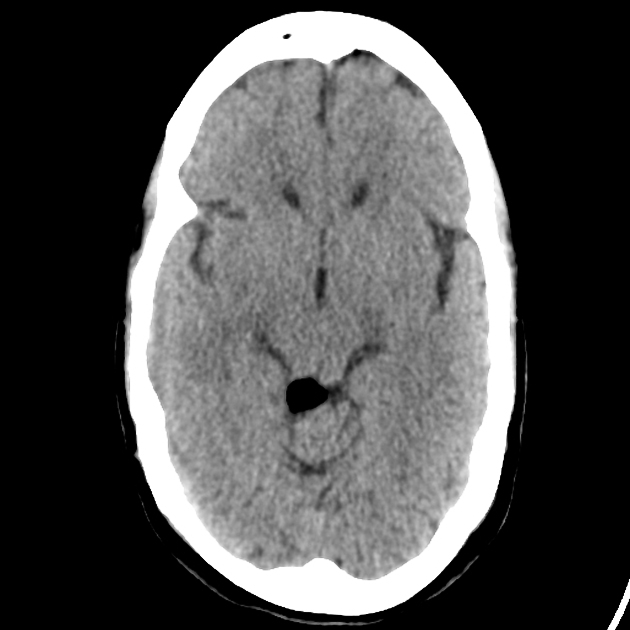
Fat-density lesion in right lower part of tectum.
From the case:
Quadrigeminal cistern lipoma











Show annotations
Download
Info
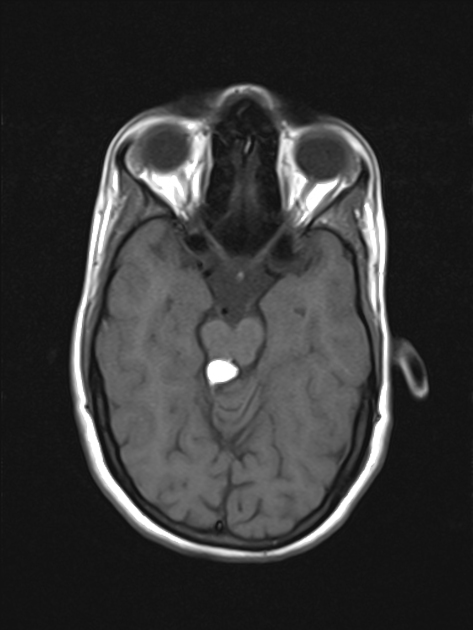
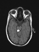
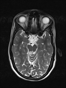
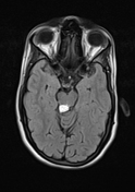
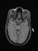
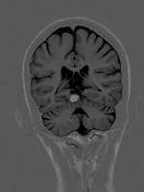
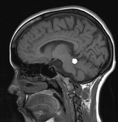
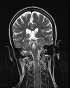
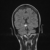
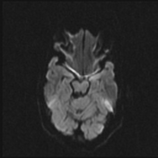
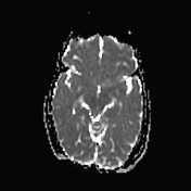
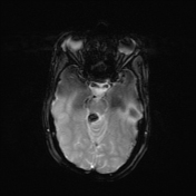
Well-defined abnormal fat-intensity lesion (13 mm x 10 mm) involving the right lower part of the tectum, appearing hyperintense on T1 and T2-weighted sequences and hypointense on T1 fat-saturated and gradient sequences.
Case Discussion
Quadrigeminal cistern lipomas make up approximately 25% of intracranial lipomas and are located within the quadrigeminal cistern. Though asymptomatic in most cases, patient presented with seizures due to mass effect.


 Unable to process the form. Check for errors and try again.