
Presentation
Macroscopic hematuria. Ultrasound revealed right renal mass.
Patient Data


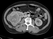
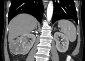
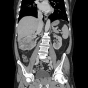
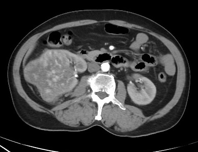
Arterially enhancing mass with areas of scattered calcification within is seen arising from the mid and lower pole cortex of the right kidney. It shows large central non enhancing areas, associated with fat stranding in perirenal space. It is causing blunting of the calyceal system. Patent renal arterial and venous vasculature. No significant perirenal retroperitoneal lymph nodes. No bony lesions. No left renal mass lesions.
Case Discussion
Renal cell carcinomas tend to present very late, unless frank hematuria begins. The fact that the kidney is a retroperitoneal organ allows lesions to take their time and grow into much large masses before becoming clinically evident. In our center. In annual screening in the last year, we detected three cases of renal cell carcinoma with lesions measuring < 3cm in size in clinically occult patients. In this case, according to CT findings, there is no evidence of locoregional spread which favors resection and possible favorable outcome.


 Unable to process the form. Check for errors and try again.