
Presentation
Work up for hematuria.
Patient Data


A low-attenuation hepatic lesion, measuring 18 mm in diameter is depicted, located at 6th hepatic segment (Couinaud`s nomenclature), revealing early peripheral, nodular enhancement and centripetal filling inferring hemangioma. Additionally, a few small cysts are scattered at liver less than 7 mm.
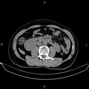
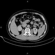
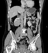
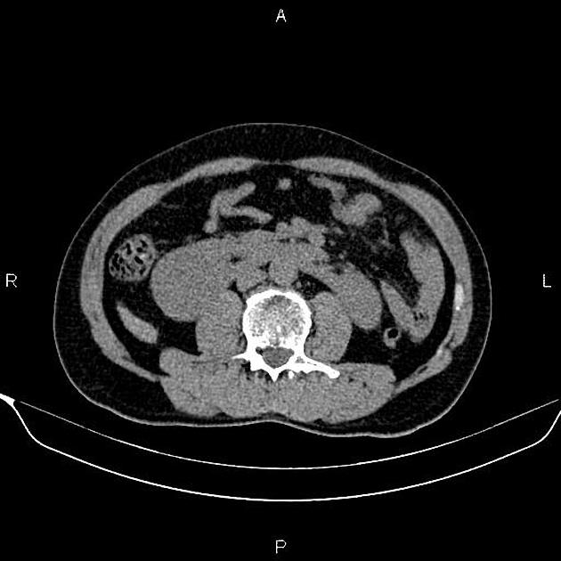
The lower poles of both kidneys are seen pointing medially and seen fused across the midline by an isthmus of functioning renal tissue with anteriorly malrotated hila denoting horseshoe kidneys.
A 53 x 51 mm mass is seen in the lower pole of right kidney. The neoplasm has a similar density to normal renal parenchyma on non-enhanced CT scan. After IV contrast media injection, the attenuation value increased from 32 HU to 78 HU. Nephrographic-phase CT scan shows that the mass enhances less than normal renal parenchyma.
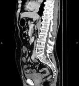
The prostate gland is enlarged and its central portion protrudes cephalad into the urinary bladder base.
Degenerative changes as osteophytosis are seen at the lumbar spine.
Case Discussion
Pathology proved renal cell carcinoma in horseshoe kidneys. CT is frequently used to both diagnose and stage renal cell carcinomas.


 Unable to process the form. Check for errors and try again.