
Presentation
Respiratory distress.
Patient Data
Age: 9 months
Gender: Male
From the case:
Respiratory syncytial virus
Download
Info
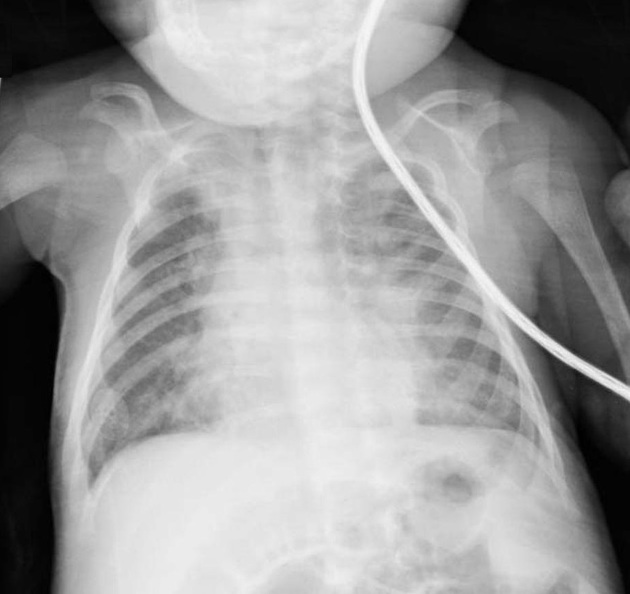
Bilateral patchy consolidation in a predominantly perihilar distribution extending to the bases. Left lower lobe and right middle lobe confluent consolidation.
In the presence of infective symptoms and respiratory distress features of syncytial virus pneumonitis (RSV).
Case Discussion
Radiograph of a patient with respiratory syncytial virus pneumonitis.
These images are from Dr. Paula Brill's excellent pediatric radiology collection.
Dr. Brill is a professor in the department of radiology (pediatric section) at Weill Cornell.
This case was donated to Radiopaedia.org by Radswiki.net.


 Unable to process the form. Check for errors and try again.