
Presentation
Unable to walk after 2 years. Short stature Fall 3 years ago. Abdominal pain.
Patient Data
Age: 5 years
Gender: Female
From the case:
Rickets


Download
Info
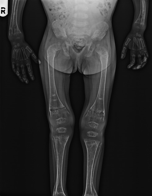
Extensive osteoporosis, remodeling, cupping, fraying and bone age nearly less than 2 years.
Case Discussion
This case depicts the classical imaging findings in active rickets.
- metaphyseal fraying and cupping in knee, shoulder, elbow and wrist joints
- widening of growth plate
- generalized osteoporosis
- delayed bone age
- widening of anterior end of ribs (rachitic rosary)
- coxa vara deformity


 Unable to process the form. Check for errors and try again.