
Presentation
Right hilar mass seen on thoracic spine series obtained to evaluate back pain.
Patient Data
Age: 35 years.
Gender: Female
From the case:
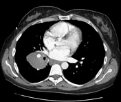
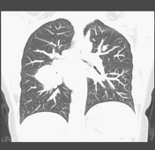
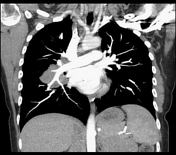
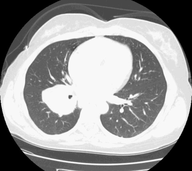
Right hilar mass
Show annotations
Download
Info

Right hilar mass, mild patchy atelectasis, otherwise normal.


Case Discussion
Because the right pulmonary artery is "silhouetted" by the mass, the mass must be in the right hilum. The hilar location of the mass is confirmed on the CT scan. This case is meant as a companion to another case with the "hilum overlay" sign, where the pulmonary artery remains visible "through" the mass because the mass is either in front of the hilum, not in the hilum. The mass was biopsied: the final pathologic diagnosis was benign reactive lymph node.


 Unable to process the form. Check for errors and try again.