
Patient Data
Age: 6 years
Gender: Male
From the case:
Right upper lobe consolidation
Download
Info
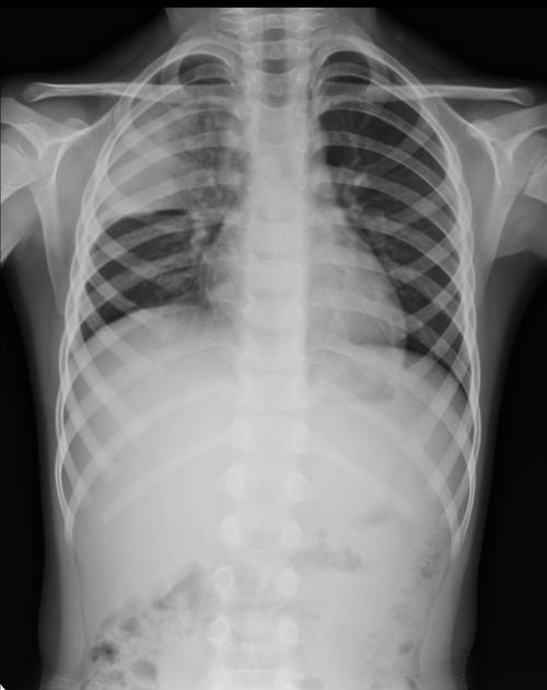
Dense consolidation peripherally in the right upper lobe.
From the case:
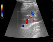
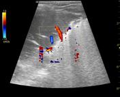
Right upper lobe consolidation
Download
Info

Airspace filling demonstrated with branching echogenic foci representing air bronchograms. The consolidated lung gives a similar appearance to the liver parenchyma.
Case Discussion
6 years old boy with reduced air entry on the right side. Air bronchograms are demonstrated on the chest radiograph and ultrasound.


 Unable to process the form. Check for errors and try again.