
Presentation
Follow-up imaging for a patent with dyspnea and recurrent pleural effusion. He underwent surgical management of aortic dissection 2 years ago.
Patient Data

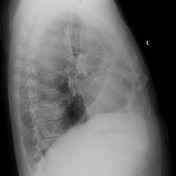
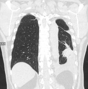
Left-sided pleural fluid collection.
Irregular area of opacity adjacent to the pleura in the left lower zone.
Reduced volume of the affected lobe and increased volume of the second lung.

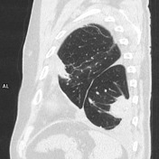

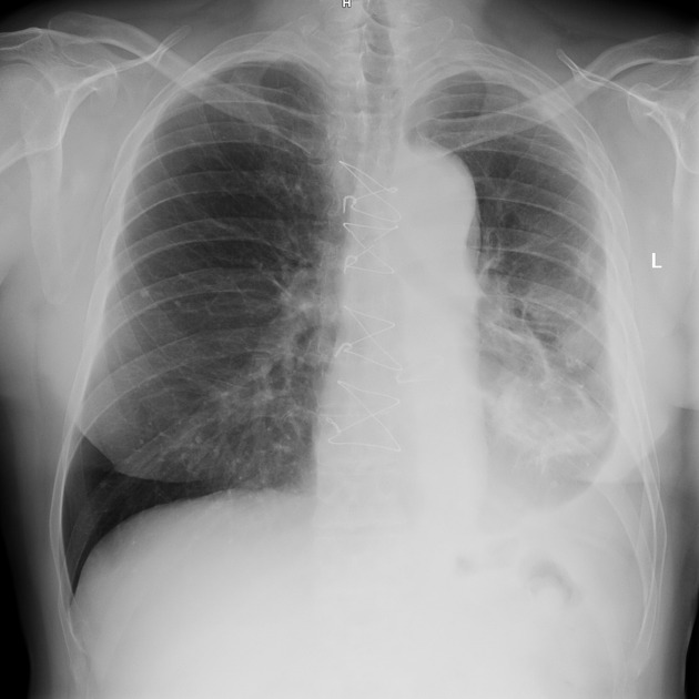
Left-sided pleural fluid collection.
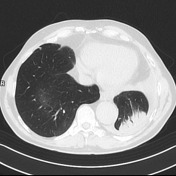
Round soft tissue density with small calcifications in the left lower lobe adjacent to the pleura with curvilinear extensions (comet tail sign) demonstrates a typical parenchymal enhancement. Subpleural location, acute angle between the mass and the pleura.
Linear bands radiating from a mass into adjacent lung tissue resembling the feet of a crow.
Reduced volume of the affected lung and increased volume of the second lung.
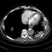
Additionally visible aortic dissection
Case Discussion
This case demonstrates all typical features of round atelectasis but differential diagnosis between other causes of lung mass can often be difficult.


 Unable to process the form. Check for errors and try again.