
Presentation
Sudden severe upper abdominal pain.
Patient Data


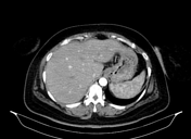
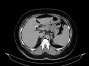
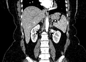
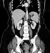
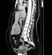
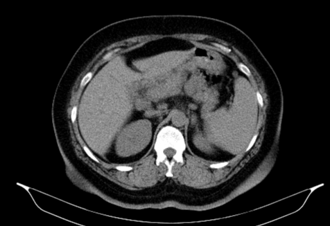
A large well defined lobulated mass is seen arising from the undersurface of left hepatic lobe segment III measuring 15cmx9.5cmx13.2cm. It shows heterogenous post contrast enhancement in the arterial phase with relative washout in the Porto venous and delayed phases. It shows internal cystic component. A large focal area of heterogenous hyperdense content is seen within the lesion at its right lateral caudal part representing hematoma. The claw sign is demonstrated particularly in coronal and sagittal planes.
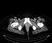
Mild dense pelvic peritoneal free fluid most likely representing mild hemoperitoneum.
Cholecystectomy clips are noted.
The patient went on to have a resection.
Macroscopic pathology
Hepatic tissue measured 14x12x3.5cm. Serialling revealed well defined mass measured 12x10x3cm, with hemorrhagic yellowish rubbery to soft cut section showing cystifications, located 0.3cm away from the least hepatic surgical margin. Rest of cut section was unremarkable.
Histology
Sections examined from specimen received revealed liver tissue showing a fairly defined tumor formed of trabeculae of bland hepatocytes showing regular nuclei with marked steatosis and intervening scattered isolated arteries.
There is focal central hemorrhage.
No regularly scattered portal areas.
Reticulin stain showing thin fibers surrounding thick and thin plates.
No malignancy.
Final diagnosis
Hepatocellular adenoma with focal hemorrhage
Case Discussion
A case presented with sudden severe upper abdominal pain shows large left hepatic lobe exophytic mass, pathologically proven to be ruptured hepatic adenoma.


 Unable to process the form. Check for errors and try again.