Presentation
Chest pain and severe dyspnea.
Patient Data


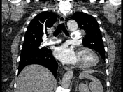
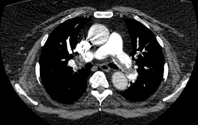
Large intra-luminal filling defect (saddle thrombus) straddles the pulmonary artery bifurcation and extends into the main pulmonary arteries, partially occluding their lumens. The thrombus continues to extend through segmental and subsegmental branches of the upper and lower pulmonary lobar arteries in both lungs, nearly occluding their lumens.
Dilated main pulmonary trunk measuring 3.6 cm in diameter, enlarged right ventricle with RT/LT ventricular diameter ratio >1, in keeping with pulmonary hypertension and right heart strain.
Case Discussion
Imaging features are consistent with saddle pulmonary embolism, which straddles the bifurcation of the pulmonary trunk, extending into the left and right pulmonary arteries. It is associated with pulmonary hypertension and right heart strain.


 Unable to process the form. Check for errors and try again.