
Presentation
Initially presented to GP with increasing shortness of breath.
Patient Data
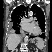


CT chest with IV contrast.
Multiple nodular lesions within the right hemithorax in the paramediastinal, peridiaphragmatic and paravertebral spaces. The largest lesion to the right of the trachea measures 35 x 20mm.
Right sided loculated pleural effusion. Pleural drain posteriorly on the right.
Case Discussion
A 100 year old female presented to her GP with shortness of breath. Chest x-ray revealed a right-sided pleural effusion of unknown etiology.
Of note initial imaging prior to further investigation was felt to be consistent with pleural mice.
A pleural drain was inserted. Pleural fluid was consistent with exudate. Microscopy showed isolated mesothelial cells, macrophages and numerous lymphocytes. Findings were negative for pulmonary malignancy and non-specific.
The lesion between the 9th and 10th ribs was biopsied.
Histology results were: fibrous tissue consistent with pleura, infiltrated by a sarcomatoid neoplasm. The tumor consists of a haphazard proliferation of variably elongated plump spindle cells with large, markedly pleomorphic nuclei showing hyperchromasia and prominent nucleoli.
History, imaging and histopathology were all indicative of sarcomatoid mesothelioma.


 Unable to process the form. Check for errors and try again.