Presentation
Discovered in a workup for osseous metastatic disease of unknown primary.
Patient Data


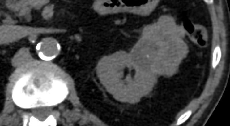
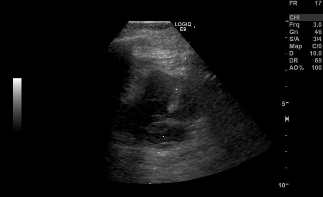
Irregular exophytic renal mass, extending off the superolateral aspect of the kidney. It is mildly hyperattenuating relative to the kidney and contains a small focus of calcification. There are regions of hypoattenuation that are compatible with necrosis. The solid component is hypoenhancing relative to the renal parenchyma. There is questionable involvement of the splenic flexure of the colon.

Ultrasound-guided core biopsy (20 gauge) of the periphery of the mass.
Case Discussion
Sarcomatoid renal cell carcinoma (sRCC) is not currently thought to represent a distinct histologic subtype of renal cell carcinoma (RCC), but represents a "final common dedifferentiation pathway." It occurs in ~16% of advanced RCCs.
sRCC develops out of advanced RCC and typically presents as a large necrotic mass with >50% having metastatic disease at presentation.
Imaging appearance of sRCC is nonspecific and there are few series in the literature attempting to characterize it.
The histologic distinction between sarcomatous RCC and advanced nonsarcomatous RCC is important for prognostic purposes since sRCC is a very aggressive variant and has a poorer prognosis. The original histologic subtype in this particular case of sRCC is unknown.


 Unable to process the form. Check for errors and try again.