
Presentation
Right wrist pain with history of a fall few months ago.
Patient Data
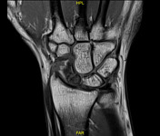

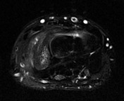
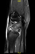

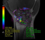

There is a complete non-union transverse fracture line seen at the junction of the proximal 1/3 and distal 2/3 of the scaphoid bone. The scaphoid bone shows increased signal on T2 denoting underlying edema. The distal segment of the scaphoid shows patchy areas of reduced signal on T1, while the proximal segment shows diffuse low signal. Following contrast administration; the distal segment shows continuous contrast uptake with variable enhancement as evident on the color-coded map, while the proximal segment shows no contrast uptake as evident by the homogeneous low signal following contrast administration, absence of color saturation and the flat dynamic curve.
Case Discussion
Avascular necrosis (AVN) is a known complication of scaphoid non-union fracture. MRI can help diagnose scaphoid AVN before the development of scaphoid collapse. The use of post-contrast MRI may not be required as the presence of decreased signal on T1 unenhanced MRI correlates well with AVN.


 Unable to process the form. Check for errors and try again.