
Presentation
Pain right groin and hemiscrotum. History of left testicular seminoma 20 years ago.
Patient Data

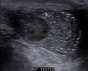
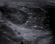
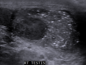
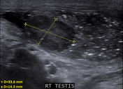
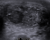
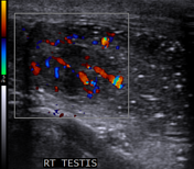
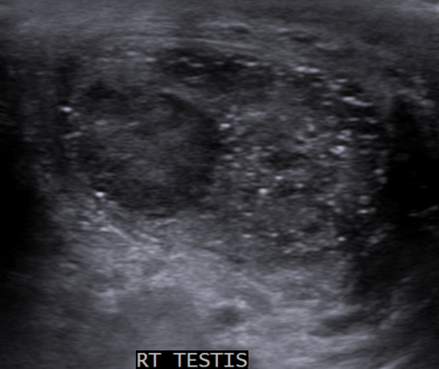
Average size right testis containing multiple microcalcifications (microlithiasis). A well defined mass measuring 1.4 x 3.3 cm is seen along the upper pole of the testis. Color Doppler ultrasound shows significant vascularity within the mass.
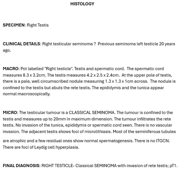
Histopathology report of the right orchiectomy specimen showed classical seminoma.
Case Discussion
Although more than 50% of the men with testicular germ cell tumor (TGCT) have testicular microlithiasis (TM), it is also commonly seen in the healthy men without any testicular malignancy. Hence a direct relationship between these two entities is quite controversial. However, this finding is significant in our case because of past history of seminoma in the contralateral testis.


 Unable to process the form. Check for errors and try again.