
Presentation
Pacemaker placement, rule out pneumothorax.
Patient Data


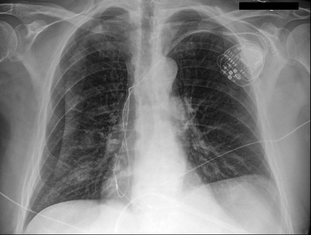
Pacemaker appropriately positioned, right-sided skinfold mimics pneumothorax. Lungs clear.
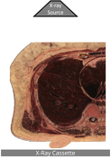
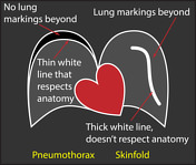
The first image shows how a skinfold may occur when the technologist slides the X-ray cassette behind a supine patient when obtaining a portable CXR: the cassette may heap up redundant skin and subcutaneous tissues. The second image shows some of the differences between a pneumothorax and a skinfold, as outlined in the discussion below.
Illustration created by Stefan Tigges
Image credit: Visible Human Project
For full attribution, see case discussion.
Case Discussion
A skinfold may be mistaken for pneumothorax, but there are differences. A pneumothorax must conform to the shape of the pleura (it won't extend outside the chest or to the contralateral side), contains only air (it will be black with no lung markings), and is a thin white line (skinfolds tend to be thicker lines). In this case, the skinfold line extends from the right hemithorax to the left hemithorax, there are lung markings beyond the skinfold line and the skinfold line is thicker than the pencil-thin pleura. If you look carefully, you can even see a second skinfold.
Attribution
Author: Visible Human Project
Original file: https://data.lhncbc.nlm.nih.gov/public/Visible-Human/Female-Images/index.html
License: The Visible Human Project was produced by the United States National Library of Medicine, National Institutes of Health and is in the public domain: https://healthdata.gov/dataset/Visible-Human-Project/krti-uwg9/data
Modifications: Square crop and creation of the skinfold in photoshop.


 Unable to process the form. Check for errors and try again.