
Presentation
Right iliac fossa pain. Blood tests revealed an elevated white blood cell count and increased percentage of neutrophils.
Patient Data
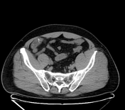

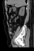
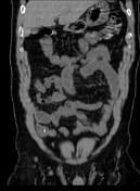
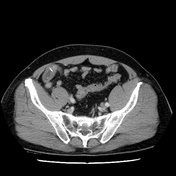
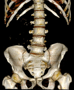
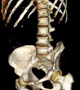

A rod-shaped structure approximately 2 cm long and 3 mm thick, with pointed ends, was located within the small bowel in the right iliac fossa. One pointed end was embedded in the bowel wall, causing thickening and edema of the corresponding bowel segment, with mild surrounding fat infiltration.
No free air or fluid was noted in the abdominal cavity. No abnormalities were observed in other organs.
Surgical report:
the ileal loops in the right iliac fossa were inflamed and congested. A sharp foreign object was observed penetrating the intestinal wall, with fibrinous exudates surrounding the perforation site
examination of the ileal loop revealed a sharp, horizontally positioned foreign object penetrating through both walls of the ileum, approximately 140 cm from the ileocecal junction
the sharp foreign object, identified as a fish bone, was removed from the ileum. Both perforation sites in the ileum were inspected. The perforations were closed with X-shaped sutures using Vicryl
Case Discussion
The imaging and surgical findings are consistent with small bowel perforation caused by a fish bone.


 Unable to process the form. Check for errors and try again.